@goodmedicine5056
General view of mitochondria ( cell biology)
Mitochondria are present in all cells except red blood cells and terminal keratinocytes.
The number, shape, and internal structure of mitochondria are often characteristic for specific cell types. When present in large numbers, mitochondria contribute to the acidophilia of the cytoplasm because of the large amount of membrane they contain. Mitochondria may be stained specifically by histochemical procedures that demonstrate some of their constituent enzymes, such as those involved in ATP synthesis and electron transport.
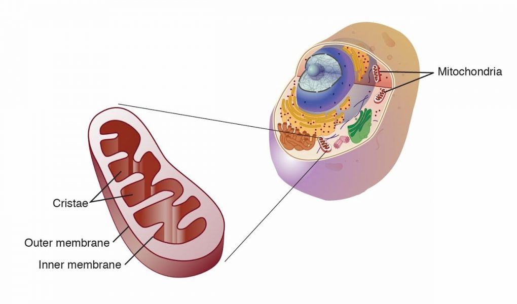
Mitochondria possess two membranes that delineate distinct compartments.
Mitochondria display a variety of shapes, including spheres, rods, elongated filaments, and even coiled structures. All mitochondria, unlike other organelles described above, possess two membranes . The inner mitochondrial membrane surrounds a space called the matrix. The outer mitochondrial membrane is in close contact with the cytoplasm. The space between the two membranes is called the intermembrane space. The following structural components of mitochondria possess specific characteristics related to their functions.
• Outer mitochondrial membrane. This 6- to 7-nm-thick smooth membrane contains many voltage- dependent anion channels (also called mitochondrial porins).
These large channels (approximately 3 nm in diameter) are permeable to uncharged molecules as large as 5,000 daltons. Thus, small molecules, ions, and metabolites can enter the intermembrane space but cannot penetrate the inner membrane. The environment of the intermembrane space is therefore similar to that of cytoplasm with respect to ions and small molecules. The outer membrane possesses receptors for proteins and polypeptides that translocate into the intermembrane space. It also contains several enzymes,
including phospholipase A2, monoamine oxidase, and acetyl coenzyme A (CoA) synthase.
• Inner mitochondrial membrane. The TEM reveals that this membrane is thinner than the outer mitochondrial membrane. It is arranged into numerous cristae (folds) that significantly increase the inner membrane surface area
(see Fig. 2.37). These folds project into the matrix that constitutes the inner compartment of the organelle. In some cells involved in steroid metabolism, the inner membrane may
form tubular or vesicular projections into the matrix. The inner membrane is rich in the phospholipid cardiolipin.
which makes the membrane impermeable to ions. The membrane forming the cristae contains proteins that have three major functions: (1) performing the oxidation reactions of the respiratory electron-transport chain, (2) synthesizing ATP, and (3) regulating transport of metabolites into and out of the matrix. The enzymes of the respiratory chain are attached to the inner membrane and project their heads into the matrix. With the TEM, these enzymes appear as tennis racquet–shaped structures called elementary particles. Their heads measure about 10 nm in
diameter and contain enzymes that carry out oxidative phosphorylation, which generates ATP.
• Intermembrane space. This space is located between the inner and outer membranes and contains specific enzymes that use the ATP generated in the inner membrane. These enzymes include creatine kinase, adenylate kinase, and cytochrome c. The latter is an important factor in initiating apoptosis . Matrix. The mitochondrial matrix is surrounded by the inner mitochondrial membrane and contains the soluble enzymes of the citric acid cycle (Krebs cycle) and the enzymes involved in fatty-acid -oxidation. The major products of the matrix are CO2 and reduced NADH, which is the source of electrons for the electron-transport chain.
https://www.facebook.com/abdullah.ashameri?mibextid=ZbWKwLرابط الحساب فيس بوك
حساب القناة تلجرام
https://t.me/abdullahmohameda
source