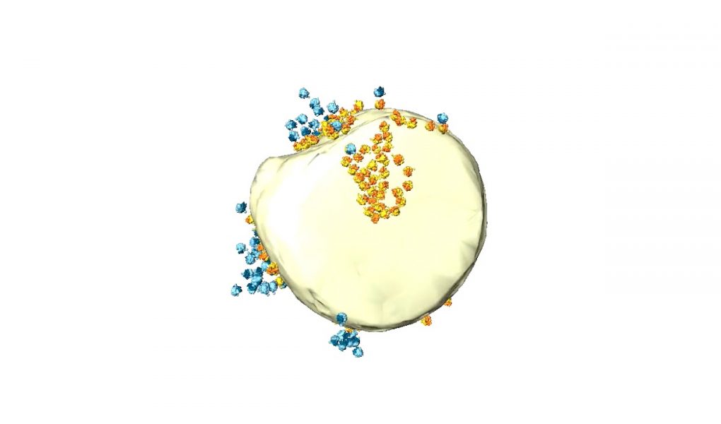
The outer membrane (yellow) and a crista membrane (transparent) are shown relative to the position of MAR-M (60S subunit yellow, 40S subunit orange) and MAR-P (60S subunit light blue, 40S subunit dark blue). Note that not all crista membranes are shown in the segmentation.
Visualization of cytosolic ribosomes on the surface of mitochondria by electron cryo‐tomography
Vicki AM Gold, Piotr Chroscicki, Piotr Bragoszewski, Agnieszka Chacinska
EMBO reports (2017)
DOI 10.15252/embr.201744261 | Published online 21.08.2017
source