
In this episode, radiologist/engineer, Raj Attariwala, explains how he was able to apply his engineering background to create a unique MRI scanner that is capable of constructing whole-body images with a resolution that is unmatched in the industry. Peter and Raj discuss the implications of such a robust, radiation-free imaging tool on the early detection of cancer. They dive deep into cancer screening and define terms such as sensitivity and specificity that are necessary to really understand this complex space. They then describe the biggest risks involved in this type of screening (false positives) and how Raj’s unique technology and process might drive down this risk substantially. But before that, they discuss all the common imaging technology from X-ray, to CT scan, to PET scans, to ultrasound, to MRI, and more. They touch on the history of each, how they work, the usefulness and limitations of each of them, as well as the varying risks involved such as radiation exposure. If you are interested in cancer screening and/or you’ve ever wondered how any radiology tool works, this episode is for you.
Subscribe on: APPLE PODCASTS | RSS | GOOGLE | OVERCAST | STITCHER
We discuss:
- Raj’s road from engineering to radiology [7:45];
- How X-ray works, the risk of radiation exposure, and the varying amounts of radiation associated with the different imaging technologies [18:00];
- Computed tomography scans (CT scans): The history of CT, how it works, and why we use contrast [27:45];
- Ultrasound: Benefits and limitations, and a special use for the heart [40:45];
- Detecting breast cancer with mammography: When is works, when you need more testing, and defining ‘sensitivity’ and ‘specificity’ [51:15];
- Magnetic resonance imaging (MRI): How it works, defining terms, and looking at the most common types of MRI [1:03:45];
- Brain aneurysms: Using MRI to find them and save lives [1:23:45];
- Raj’s unique MRI technology [1:30:00];
- The risk of false positives in cancer detection, and how Raj’s MRI can reduce the number of false positives (i.e., increase specificity) [1:43:40];
- The unique software Raj created to pair with his MRI machine [1:51:15];
- Comparing the radiation exposure of a whole-body PET-CT to Raj’s equipment (DWIBS-MRI) [1:53:40];
- How diffusion-weighted magnetic resonance imaging (DW-MRI) has revolutionized cancer screening [1:55:15];
- Why a DW-MRI is still not a perfect test [1:59:00];
- The potential for advancing MRI technology: Where does Raj think it could improve in the next 5-10 years? [2:03:00];
- Are there any commercially available scanners that can match the resolution of Raj’s images? [2:06:00];
- Machine learning: When and where might machine learning/AI impact the field of radiology? [2:08:40]; and
- More.
§
Raj’s road from engineering to radiology [7:45]
How they met
- Peter and Raj met through a mutual friend that told Peter he had to go and check out Raj’s fancy MRI machine
- Peter flew to Raj’s clinic in Vancouver and got a full-body MRI in 2015 for the first time
*See Peter’s instagram post showing video of the scan*
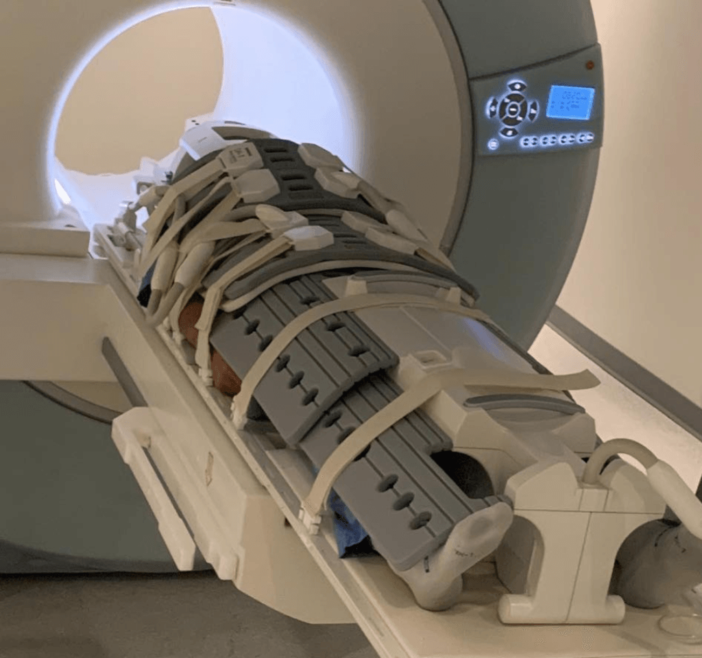
Figure 1. Peter going into Raj’s Prenuvo MRI machine.
Raj’s business
- AIM Medical Imaging is a private scanning company which Raj started to be able to “play” with his MRI technology
- His clinic is called Prenuvo which was started in order to “put the power of preventive medicine into a patient’s hands”
Raj’s road to radiology
- Raj got a chemical engineering degree
- during that period of time I realized that I actually like the body and physiology and how that works
- Then decided to get a master’s and PhD in biomedical engineering at Northwestern
- During that time, he and his team built a robot that could do keyhole surgery in the eye that wound up attracting a lot of attention from physicians from top-tier universities
- The desire to be able to speak the language of doctors, Raj decided to go to medical school
Med school
- “Hated every minute of it”
- Raj likes answers to questions and soon realized that there’s a lot of things in the body, and physiology, and pathophysiology that we just don’t fully understand
“What I would actually start to boil it down to is that I would actually go back to my engineering pathophysiology texts, and I would actually read them and talk to the PhD guys, and they would actually give me the theories on what they thought was happening. When you actually got that theory, it was almost like planting a seed, and then you actually understood how the entire tree would look.”
Technology in medicine
- Raj’s first love was technology so he decided to put those things together
- Worked on some of the very first surgical robotic machines ever built
- But in that process, Raj realized that the best place to focus on technology in medicine was radiology (imaging technology)
Radiology
- Started in an area called nuclear medicine, which is a small specialty within radiology where you’re actually looking at functional imaging, how things work, how do things change, what happens over time
- Really liked nuclear medicine because it was showing you what’s happening when things are normal and then when things become abnormal (Raj particularly loved how the answer was black and white which is rare in medicine)
Analogy
- Radiology is like anatomy, a blueprint… where you actually see what’s going on, you actually see the changes, and you see the shapes of things, and you use that very much as a blueprint for a building to actually see what it does
- Whereas nuclear medicine, instead of the blueprint, you actually know there’s all these people carrying letters, moving in and out of this building (We don’t really know where the building walls are, but we know that there must be something happening there)
- When you put the blueprint together with the movement inside the area of interest, you start to realize “oh, these are postal workers carrying things, and here’s the geometry of the building. That power really is actually quite useful.”
1 +1 = 3
- The first device to combine functional imaging with nuclear medicine imaging was the positron-emission tomography-computed tomography (PET-CT)
- These two separate modalities of functional imaging and anatomic imaging come together to actually make something better than each part individually (i.e., 1+1=3)
Advice Peter got from radiology people while in med school:
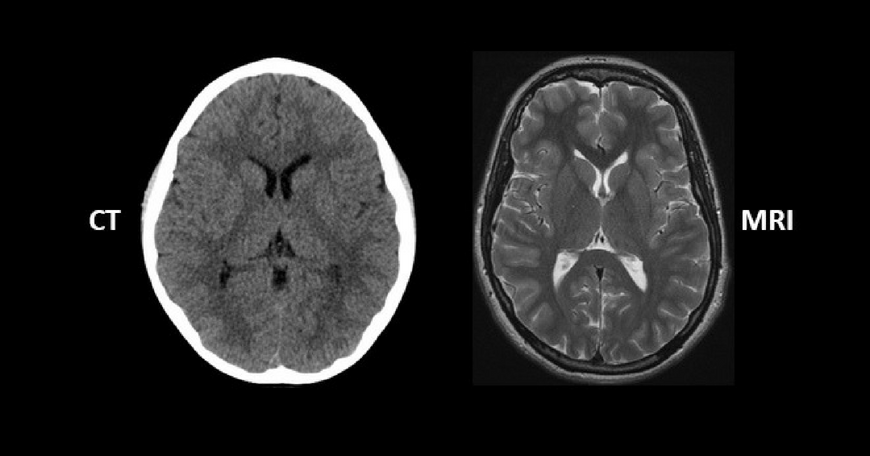
“Anytime you order a test, in the back of your mind you have to be asking yourself, ‘Do you want anatomical information, or functional information or both?’”
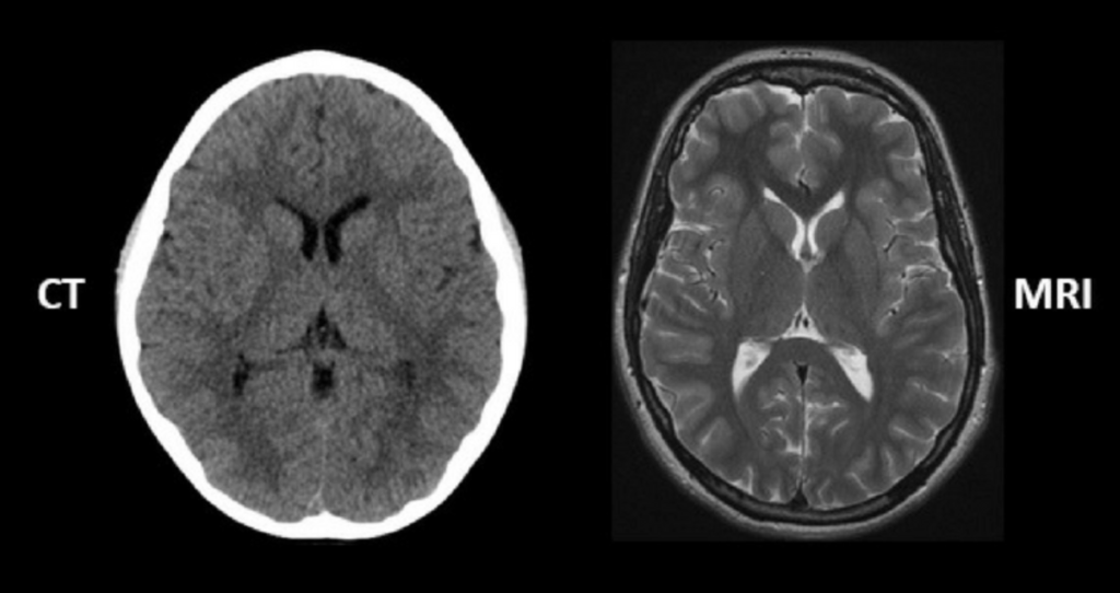
Figure 2. An example of an anatomical image of the brain with CT scan and with an MRI. Image credit: regencymedicalcentre.com
{end of show notes preview}
Rajpaul Attariwala, M.D., Ph.D.
Raj is the Medical Director at AIM Medical Imaging in Vancouver.
Over the past decade, he has been effectively creating a new way of doing MRI by fine-tuning the hardware and building unique software to create a completely revolutionary product and process by which to look at the body using the technology of magnetic resonance.
He is a dual board certified Radiologist and Nuclear Medicine physician certified in both Canada and the United States. He received his formal training at UBC with periods of specialized medical training at Memorial Sloan Kettering Cancer Centre (New York), UCLA, and USC.
Raj completed a Ph.D. in biomedical engineering at Northwestern.
