Cytoplasmic streaming can easily be observed in epidermal onion cells. Preparing microscopic slides from onion cells is fairly easy and is therefore one of the most studied specimen in schools.
With proper illumination techniques, it is possible to not only observe the very prominent cell nucleus but also many other organelles inside the cell including Leucoplasts, Spherosomes and Mitochondria.
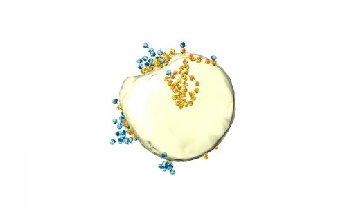
Mitochondria are particularly interesting as they are the main energy producer in the cell. However, it is rather difficult to distinguish mitochondria from other organelles using brightfield or even DIC. Fluorescence microscopy can help as dyes that specifically stain mitochondria are available. For this video, I used Rhodamine 123 which is a cationic dye that stains mitochondri based on their membrane potential and can be excited by blue light while emitting green fluorescence.
Staining procedure:
1. The Rhodamine 123 stock solution (5mg/ml) was diluted to 5ug/ml in 1x PBS in a standard 1.5ml Eppendorf tube
2. The onion epidermal cell layer was placed into the staining solution and incubated for 20min
3. The staining solution was removed and the onion cells were washed 3x with 1ml of 1x PBS
4. The onion cells were then placed on a slide, and observed under blue excitation light
I used an Olympus Vanox AHBT3 with the original blue fluorescent cube together with a Cree XM-L2 LED as the excitation light source. I used the following objectives: Olympus SPlanApo 20/0,7, SPlanApo 60/1,4, UVFL and a Leitz NPL Fluotar 50/1,0. The camera was a Panasonic S1 that is adapted using an Olympus NFK 2,5x photo eyepiece.
Background music:
Glimpsing Infinity – Asher Fulero
source